Products
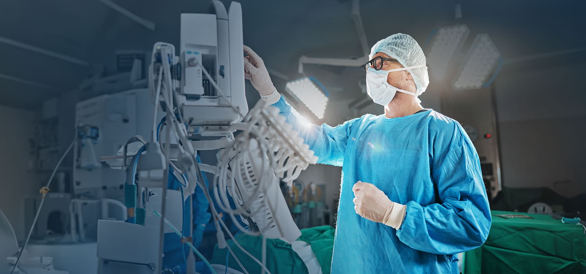
Surgical Operating Microscope

Neuro & Spine, ENT and Plastic & Reconstructive
Surgical Microscopes
Model
Ocula
Exceptional Depth Perception
Stable, Ergonomic Design
Engineered to meet the critical demands of neurosurgical and spine procedures, where precision and clarity are vital. High-definition optics, superior illumination, and smooth motorised movements allow surgeons to work with enhanced confidence and control.


Enables real-time interaction and verbal coordination between the surgeon and co-observer, improving teaching clarity and procedural teamwork.
Superior Visualisation
- - Provides high-resolution, stereoscopic magnification for intricate neural and spinal structures.
- - Enhances differentiation between healthy tissue, lesions, and critical vascular structures.
Enhanced Surgical Precision
- - Enables accurate dissection and micro-manipulation within confined anatomical spaces.
- - Facilitates delicate suturing and nerve preservation during microsurgical interventions.
Improved Depth Perception & Illumination
- - Coaxial LED or xenon illumination ensures uniform lighting without shadows.
- - Three-dimensional depth perception improves hand-eye coordination and procedural safety.
Better Ergonomics for the Surgeon
- - Adjustable viewing angles and motorised positioning reduce surgeon fatigue during long procedures.
- - Promotes steady hand posture and minimizes tremors for more controlled movements.
- - Supports image and video documentation for teaching, review, and medico-legal record.
Enhanced Outcomes & Reduced Complications
- - Greater surgical accuracy leads to minimal tissue trauma and reduced blood loss.
- - Improves postoperative recovery and reduces complication rates.
Educational & Collaborative Benefits
- - Integrated 4K ultra definition visualisation and recording options aid in training, live demonstrations, and team coordination.
- - The co-observer tube allows an assistant surgeon, trainee, or observer to view the same surgical field seen by the main surgeon in real time.
- - It provides binocular vision, maintaining the same magnification and depth perception as the primary surgeon. - Enhanced Team Coordination
- - The assistant can monitor every movement and step of the surgeon.
- - This enables better instrument handling, anticipation of needs, and smoother surgical workflow.
- - Educational and Training Purpose
- - In teaching hospitals and training centers, co - observer tubes allow residents and fellows to learn from live surgeries directly through the microscope optics.
- - Facilitates hands - on guidance, as the trainer can correct or demonstrate techniques while the trainee observes simultaneously.
- - Documentation and Demonstration
- - When coupled with a camera adapter or video system, the co - observer tube can also assist in recording or broadcasting the procedure for academic or review purposes.
- - Flexible Positioning
- - It can be mounted on either side of the microscope to suit surgeon–assistant ergonomics
- - Adjustable inter - pupillary distance ensures comfortable viewing for different users.
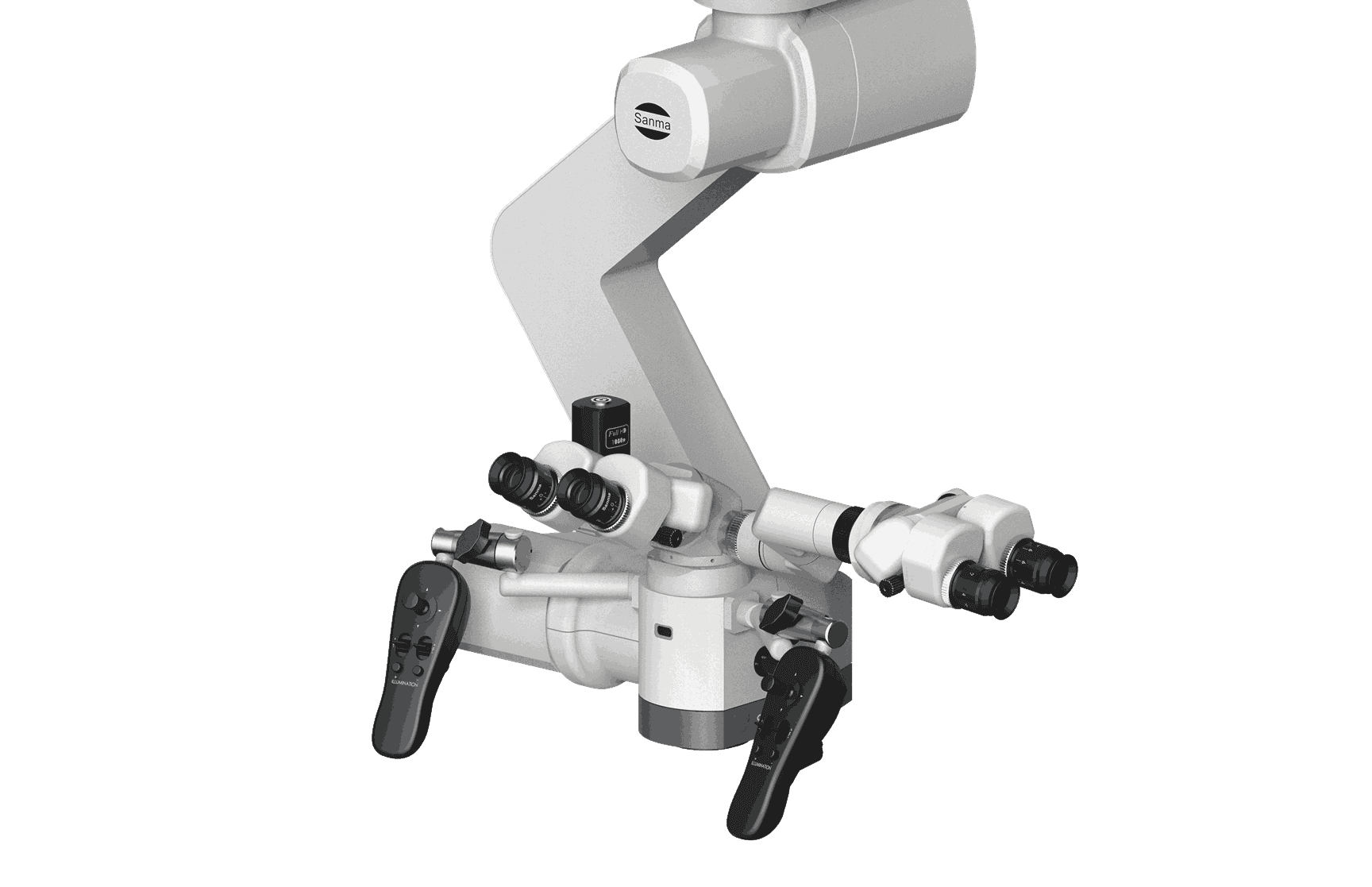
Side Co-observer tube
- - Binocular optical design with independent focusing.
- - Image parity identical to the main binoculars—so no inversion or mirror effect.
- - Optional rotatable mount for flexible positioning during multi-surgeon setups
- - 3 axis adjustable angles and image rotating